


anatomy of the chicken
next
I
II-III
IV-V
VI-VII
VIII
back


This unique presentation of color transparencies shows the fundamental structure and anatomy of the chicken, as they may be observed during a dissection study. The colors are intended to aid identification and not to represent the true colors of the organs and parts.
Originally produced by Merck Chemical Division and Merck Sharp and Dohme International in 1964.
Permission to reproduce the images obtained from Merial Australian Pty Ltd.
Let’s start
How to use this guide
Click on the numbers to see the captions. Click on the captions to close them.
Click on the ‘details images’ for an expanded view.
I

Click on green buttons for further information.


Index to plates
I. Lateral aspect of chicken
Lateral aspect of chicken (at right)
Diagram of avian eye (tap to view this page)
Skeletal system (left half)
Brain
Pulmonary system (left half)
Diagram of air sacs (ventral aspect)
Pulmonary system (left half) plus heart
Diagram of heart and great vessels (ventral aspect)
Gastrointestinal tract (left half)
Diagram of structures about the gizzard
Gastrointestinal tract (right half)
Diagram of cloaca
Genitourinary system and right half of skeleton
Diagram of oviduct
Musculature
Diagram of nephron
I
Ia
II
IIa
III
IIIa
IV
IVa
V
Va
VI
VIa
VII
VIIa
VIII
VIIIa
anatomy of the chicken
next
I
II-III
IV-V
VI-VII
VIII
back


2. anterior chamber
6. posterior chamber
10. pecten
4. scleral ring
11. optic nerve

5. lens


3. iris
1. cornea


7. retina
8. choroid
9. sclera
Ia Avian eye
1a - Tap to view
I
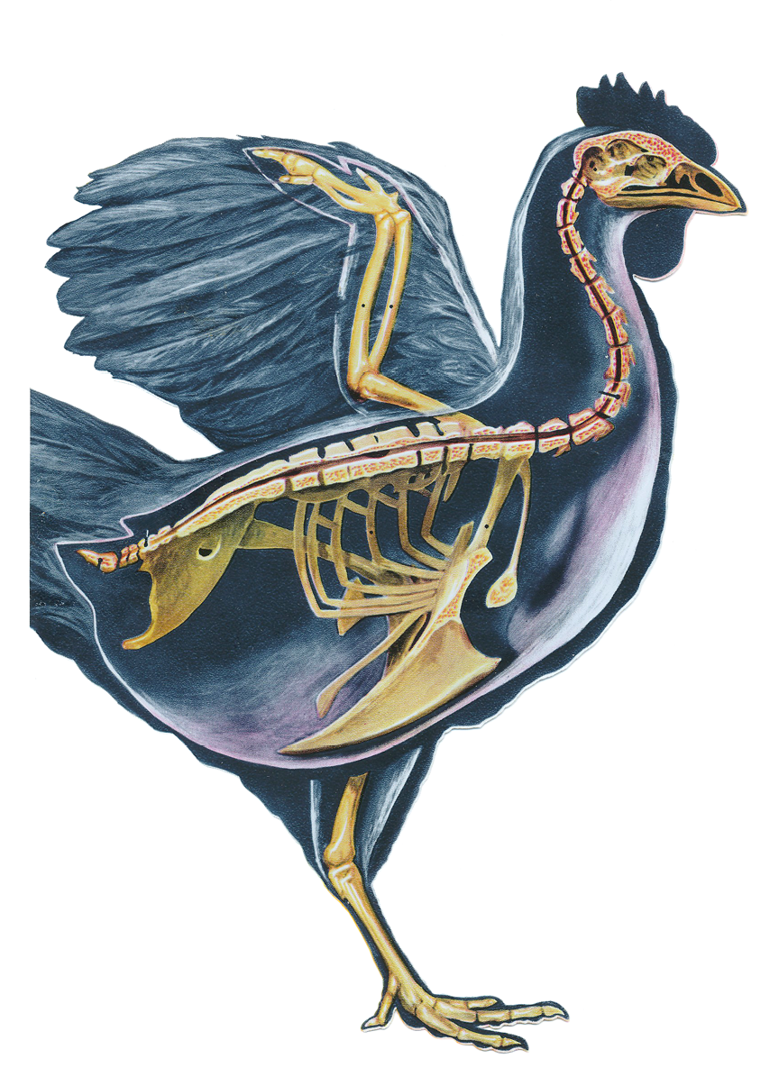
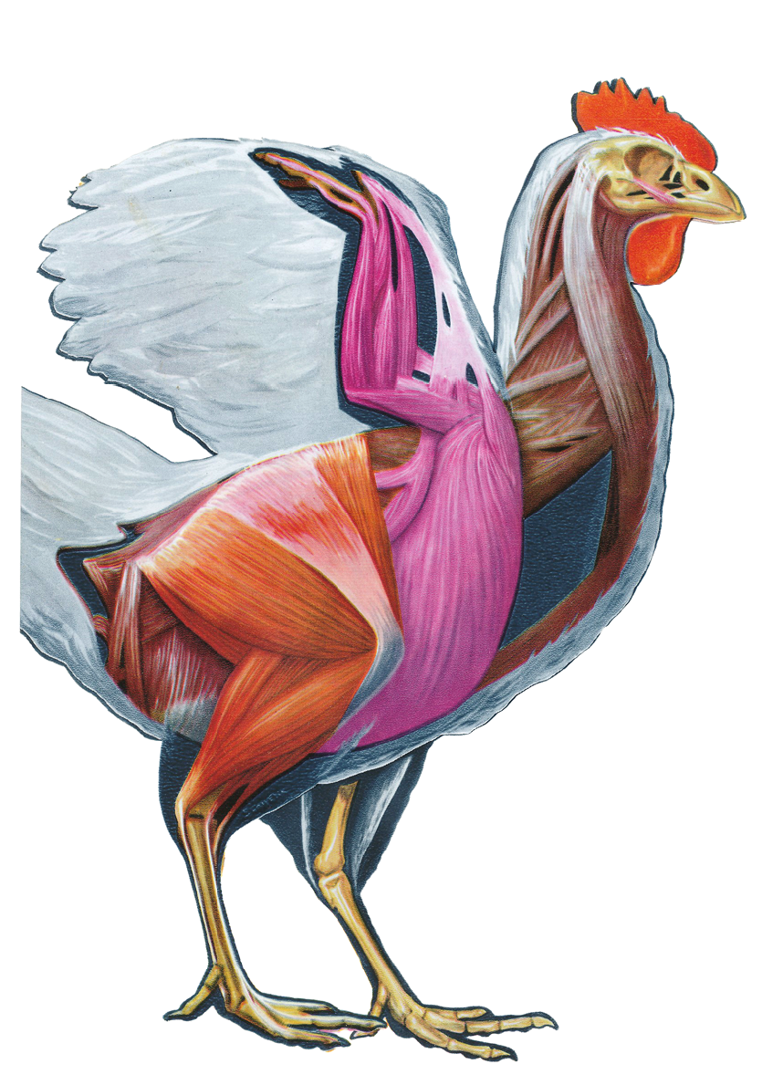
Plate I - Lateral aspect of chicken
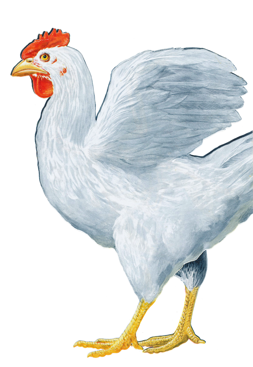
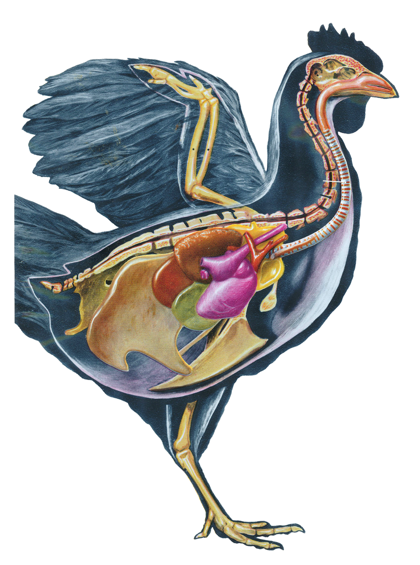
The general form of the domestic chicken is presented in a position for anatomical study. Structurally, birds share with mammals the distinction of being the most highly specialized of vertebrates.
While basically similar to mammals anatomically, evolution has brought about modifications for adaptation to flight. The forelimb is changed to a wing in which the manus has been reduced by the disappearance of digits and fusion of metacarpals.
Reduction of the manus combined with comparative simplicity of movements has removed the necessity of large and strong forearm muscles. The muscles of the pectoral region (breast), on the contrary, are powerful to propel the bird in flight and are large and well developed.
The magnitude of this development may be gathered by the fact that the muscles composing the breast weigh about as much as the all the rest of the muscles of the body together, contributing about one-twelfth of the entire body weight.
I
Ia
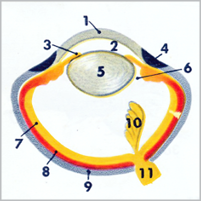
Plate Ia - Diagram of avian eye
The eye is composed of the outer, transparent cornea (1) enclosing the anterior chamber (2) and a pigmented diaphragm, the iris (3), which regulates the amount of light entering through the pupil by dilation and contraction.
The yellowish color of the iris is probably due to the presence of fat globules contained within the cells.
The scleral ring (4) and sclera (9) are dense and white while the crystalline lens (5) is a strongly biconvex transparent body suspended immediately behind the iris. The anterior chamber, enclosed in front by the cornea and behind by the iris and lens, communicates with the posterior chamber (6) through the pupil.
The chambers are filled with aqueous humor, a clear fluid about 98% water with a little sodium chloride and traces of albumin and extractives. The retina (7) or nervous tunic of the eyeball consists of nerve cells and fibers directly or indirectly continuous with the optic nerve (11). A remarkable structure known as the pecten (10) projects from the region of the entrance of the optic nerve.
Ia


II Skeletal system (left half)
III Pulmonary system (left half)
anatomy of the chicken
next
I
II-III
IV-V
VI-VII
VIII
back
Click here to view/hide focus area
12
12
skull

anatomy of the chicken

26
26
coracoid

20
20
sternum

19
19
ribs

14
14
thoracic vertebrae

28
28
femur

24
24
radius

40
40
left lung

39
39
trachea

63
63
cloaca

41
41
axillary air sac

62
62
large intestine

43
43
abdominal sac

27
27
clavicle

25
25
humerus

23
23
ulna

21
21
phalanges

22
22
metacarpus

29
25
tibia

30
30
metatarsus

31
31
phalanges

42
42
anterior thoracic sac

44
44
heart

55
55
crop

17
17
pygostyle

18
18
pelvis

15
15
lumbosacral vertebrae

16
16
coccyx

13
13
cervical vertebrae

38
38
nasal cavity


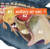
IIIa Diagram of air sacs (ventral aspect)
41. axillary air sac
40. left lung
42. anterior thoracic sac
46. posterior thoracic sac
45. syrinx
43. abdominal sac


IIIa: Tap to view
IIa: Tap to view
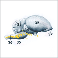

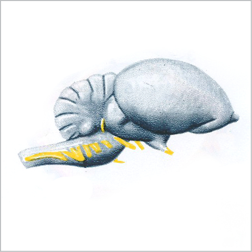
IIa Brain
32. cerebrum
37. olfactory lobe
35. medulla
33. cerebellum


34. optic lobe

36. cranial nerves (yellow)
IIa

Plate IIa - Brain
The chief structures of the brain are the large cerebrum (32) of the forebrain, the cerebellum (33) of the hindbrain and the optic lobes (34) of the midbrain.
The medulla oblongata of the hindbrain bears a close resemblance to the spinal cord with which it is continuous.
There are 12 pairs of cranial nerves (36), the first of which, the olfactory (37), forms a projection on the anterior aspect of each cerebral hemisphere.
IIa


III
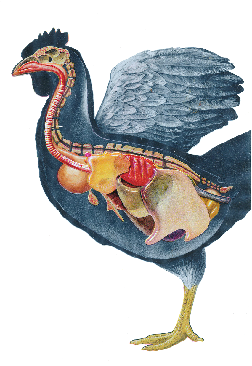
Plates III and IIIa: Pulmonary System and Diagram of Air Sacs
The nasal cavity (38) is short and narrow while the trachea (39) or windpipe is relatively long, ending by branching into the right and left bronchi.
The lungs (40) are relatively small in comparison with the size of the thorax and are closely applied to the vertebrae and ribs.
The axillary air sac (41) is connected with the cranial end of the lung transmitting air to the sternum, sternal ribs, shoulder-girdle and humerus.
The anterior thoracic air sacs (42) stretch from the clavicular to the abdominal sacs and unlike the other air sacs do not communicate with the interior of the bones.
The abdominal air sacs (43) are large and communicate directly with the cavities of the sacrum, pelvis and femur. The heart (44) is seen in part projecting from behind the axillary and thoracic air sacs.
The syrinx (45) or broncho-tracheal larynx is demarcated by a lateral compression where the trachea divides into right and left bronchi. Elastic membranes homologous to those of the mammalian larynx are found in this organ.

III

II
II
A view of the left half of the skeleton shows the skull (12), the thirteen cervical vertebrae (13) of the neck, the seven thoracic vertebrae (14), the fused lumbosacral vertebrae (15), the five or six coccygeal vertebrae (16) of which the last, produced by the union of several vertebrae, is the largest.
The latter, known as the pygostyle (17), forms a foundation for the feathers of the tail. The pelvis (18) is formed by the union of the ileum, ischium and pubis. Of the seven pairs of ribs (19) the first and second and sometimes the seventh do not reach the sternum (20).
The phalanges (21) are rudimentary while the metacarpus (22) is in the form of a single bone produced by the union of three osseous elements corresponding to the first, second and third metacarpal bones of the mammalian limb.
The ulna (23) and radius (24) form the bones of the forearm while the stout and slightly curved humerus (25) has an ovoid head for articulation with the scapula and coracoid (26).
The clavicle (27) is thin, rodlike and slightly bent. The united clavicles form the furcula or wishbone which supports the shoulders preventing them from coming too close together during flight.
The femur (28) is stout and cylindrical while the tibia (29) with its attached, poorly developed fibula is much longer than the femur.
The adult metatarsus (30) is represented by one long bone composed of second, third and fourth metatarsal bones in union. The phalanges (31) form the four digits.


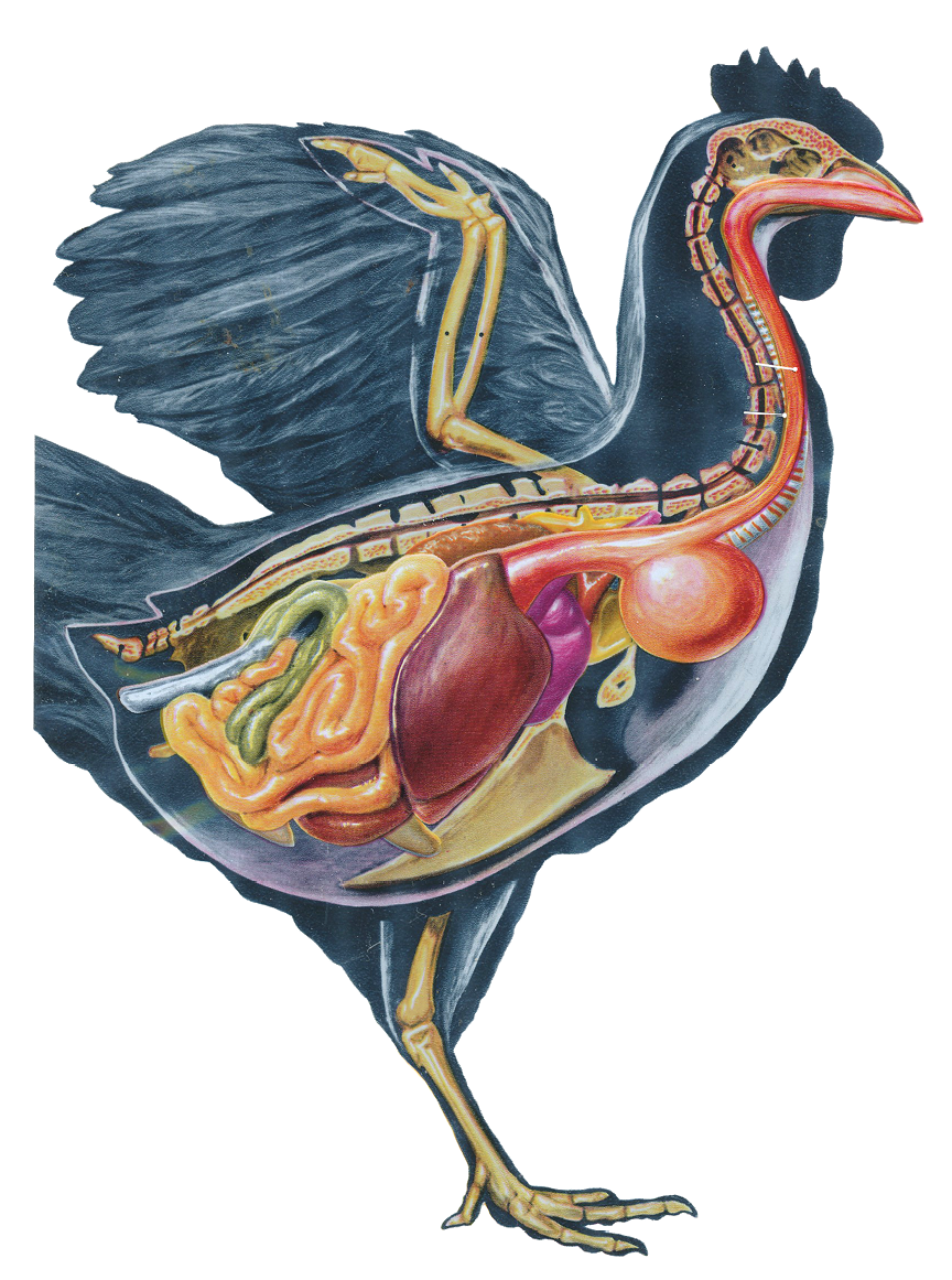
IV Pulmonary system (left half) plus heart
V Gastrointestinal tract (left half)

anatomy of the chicken
41
41
axillary air sac

39
39
trachea

20
20
sternum

24
24
radius

54
54
esophagus

63
63
cloaca

59
59
pancreas

58
58
duodenum

61
61
ceca

62
62
large intestine

57
57
gizzard

60
60
small intestine

27
27
clavicle

23
23
ulna

22
22
metacarpus

21
21
phalanges

29
29
tibia

30
30
metatarsus

55
55
crop

56
56
proventriculus

64
64
liver

17
17
pygostyle

15
15
lumbosacral vertebrae

16
16
coccyx

13
13
cervical vertebrae

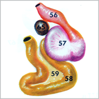

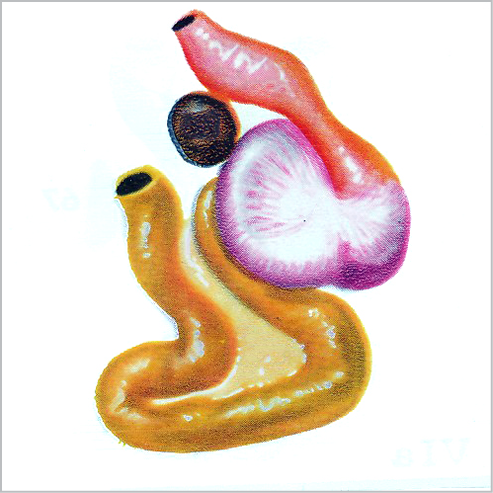
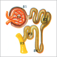
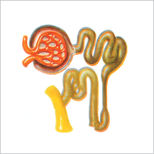
Va Diagram Structures about the gizzard
56. proventriculus
57. gizzard
59. pancreas
65. spleen
58. duodenum


Va: Tap to view
18
18
pelvis

42
42
anterior thoracic sac

46
46
posterior thoracic sac

44
44
heart

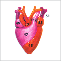

IVa Diagram of heart and great vessels
53. left atrium
47. right ventricle
49. right atrium
48. left ventricle


50. aorta


52. pulmonary artery
51. left anterior vena cava

IVa: Tap to view
anatomy of the chicken
next
I
II-III
IV-V
VI-VII
VIII
back
12
12
skull

25
25
humerus

43
43
abdominal sac


IVa
Plate IVa - Diagram of heart and great vessels
Detailed structure of the heart and associated blood vessels. The right ventricle (47) pumps blood to the lungs by means of the pulmonary artery (52) while the heavily walled left ventricle (48) forces blood to the body through the aorta (50).
The right atrium (49) receives blood from the caudal vena cava and two cranial venae cavae (51) and transmits it through a strong muscular plate (analogous to the tricuspid valve of mammals) into the right ventricle. The left atrium (53) is a common opening through which the pulmonary veins pour blood after it has passed through the lungs.
IVa


IV
Plate IV - Pulmonary System
The gross appearance of the respiratory system from the medial aspect.

IV

V

V
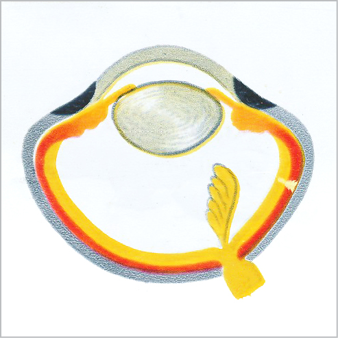
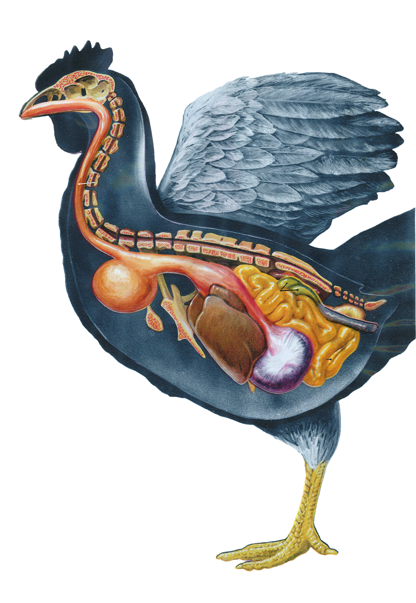
Plates V and Va: Gastrointestinal tract and Diagram of structures about the gizzard
The esophagus or gullet (54) has at the thoracic inlet a marked dilation, the crop (55) which serves to store food after rapid ingestion.
The esophagus terminates at the glandular stomach or proventriculus (56) which is relatively small but richly supplied with glands and lymphoid tissues. The muscular stomach or gizzard (57) immediately succeeds the glandular stomach from which it is separated merely by a constriction.
The small intestine (60) begins at the exit from the gizzard and is of relatively uniform calibre throughout. Of the three parts, duodenum, jejunum and ileum, only the first (58) can be distinguished.
The elongated loop of the duodenum, consisting of descending and ascending limbs, extends as far posteriorly as the entrance to the pelvis. Between the limbs of the duodenum lies the pancreas (59). The ceca (61) are blind tubes arising at the junction of the small and large intestine (62). The large intestine is very short, ending in the cloaca. Some anatomists describe the large intestine as consisting of two parts, the colon and rectum. The cloaca (63) is the common opening of the digestive and urogenital tracts. The large, dark-brown liver (64) is divided into two lobes, the right being the larger. The relatively large gallbladder lies on the right posterior part of the visceral surface of the liver. The spleen (65) is a reddish-brown rounded structure situated near the junction of the proventriculus and gizzard and is associated with the circulatory system.
31
31
phalanges


VI Gastrointestinal tract (right half)
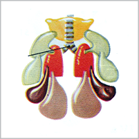
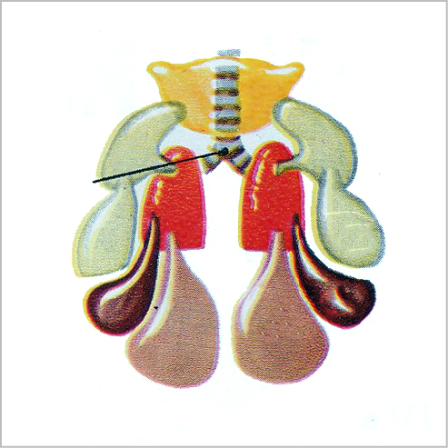
VII Genitourinary system and right half of skeleton

12
12
skull

anatomy of the chicken
20
20
sternum

24
24
radius

39
39
trachea

68
68
ovary

75
75
kidney

70
70
infundibulum

67
67
oviduct

27
27
clavicle

25
25
humerus

23
23
ulna

21
21
phalanges

29
29
tibia

30
30
metatarsus

69
69
ovum

17
17
pygostyle

15
15
lumbosacral vertebrae

16
16
coccyx

13
13
cervical vertebrae

54
54
esophagus

58
58
duodenum

61
61
ceca

60
60
small intestine

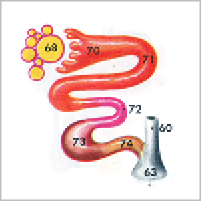

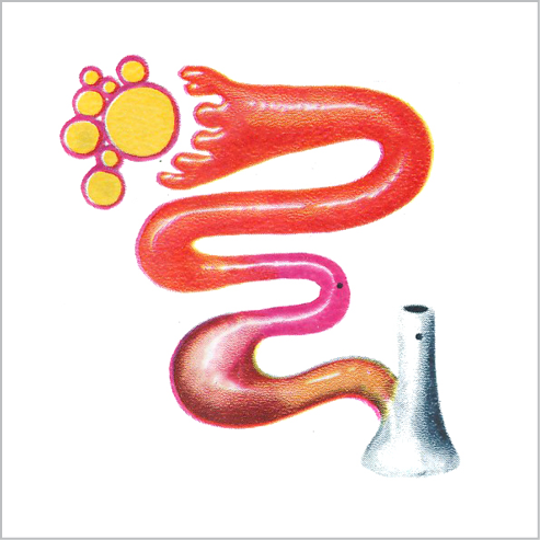
VIIa Diagram of oviduct
71. ampulla of oviduct
60. small intestine
73. uterus
68. ovary
63. cloaca


72. isthmus of oviduct

70. infundibulum

74. vagina

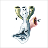

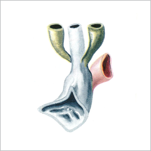
VIa Diagram of cloaca
67. oviduct
63. cloaca
73. uterus
61. ceca
66. bursa of Fabricius


60. small intestine

VIIa: Tap to view
VIa: Tap to view
22
22
metacarpus

55
55
crop

56
56
proventriculus

anatomy of the chicken
next
I
II-III
IV-V
VI-VII
VIII
back


VI
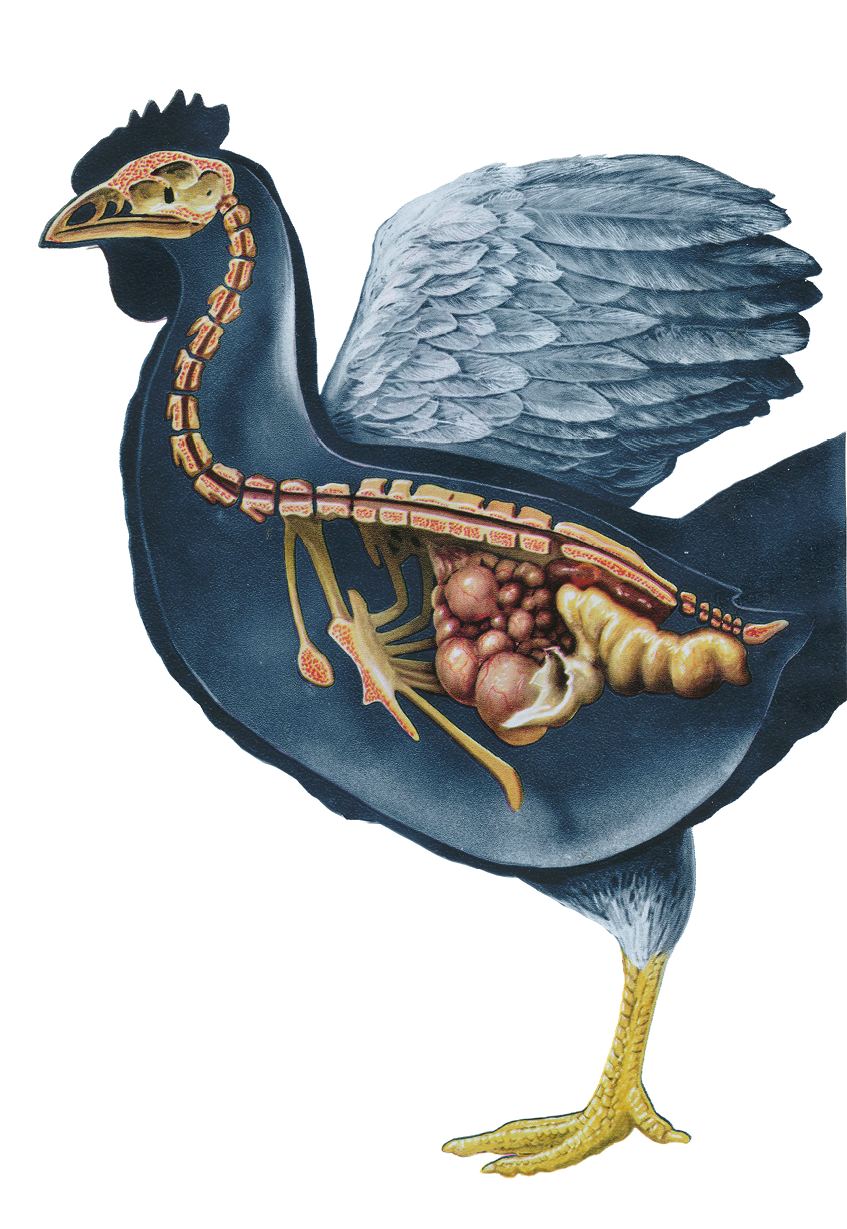
Plate VI - Gastrointestinal tract (right half) and VIaDiagram of cloaca
The terminal portion of the gastrointestinal tract viewed from the right side shows the junction of small and large intestine with entrance of the oviduct (67) and bursa of Fabricius (66). The latter is found best developed in chickens about 4 mo. of age and has usually disappeared at 1 yr.

VI

VII
Plates VII Genitourinary system and VIIaDiagram of oviduct
The ovary (68) with ova (69) in various stages of development is an unpaired structure in the hen (while there are two ovaries in the embryo the right does not develop).
The left ovary lies in the dorsal part of the abdominal cavity opposite the last two ribs. The oviduct (67) varies in appearance according to its functional state. Its anterior part corresponding to the Fallopian tube of the mammal has a slit-like opening leading to the expanded infundibulum (70) succeeded by the more narrow ampulla and isthmus.
Behind this the oviduct enlarges to form a wide, thick-walled tube (73) which may be regarded as the homolog of the mammalian uterus. This is succeeded by the narrow vagina (74) which opens into the cloaca at the left ureteral opening.
The kidneys (75) lie along each side of the vertebral column from the vertebral end of the sixth rib to the iliac fossa. Each kidney consists of three or four lobes.

VII
40
40
left lung

41
41
axillary air sac

31
31
phalanges


VIII Musculature
KEY TO IDENTIFIED ANATOMICAL ASPECTS
79
79
pelvic limb muscles

78
78
pectoral girdle muscles

29
29
tibia

30
30
metatarsus

76
76
cervical muscles

77
77
pectoral limb muscles


VIIIa Diagram of nephron
82. proximal convoluted tubule
83. distal convoluted tubule
85. collecting tubule
80. glomerular tuft
84. loop of Henle


81. Bowman’s capsule

VIIIa: Tap to view
anatomy of the chicken
next
I
II-III
IV-V
VI-VII
VIII
back

VIII
Plate VIII - Musculature
The cervical (76), pectoral limb (77), pectoral girdle (78) and pelvic limb (79) muscles of birds are highly modified to meet the needs of function in locomotion.

VIII
VIIIa

Plate VIIIa - Diagram of nephron
Microscopic appearance of the functional renal corpuscle. The glomerular tuft (80) enclosed by Bowman’s capsule (81).
The proximal convoluted tubule (82) leaves the capsule by a short neck and is connected to the distal convoluted tubule (83) by the loop of Henle (84) terminating in the collecting tubule (85).
VIIIa

31
31
phalanges
